Introduction
Triangular Fibrocartilage Complex (TFCC) injuries are a common yet frequently underdiagnosed cause of ulnar-sided wrist pain, particularly in athletes and individuals involved in repetitive wrist loading. The TFCC plays a crucial role in wrist stability, load transmission, and smooth forearm rotation. A sound understanding of its anatomy, injury patterns, and management options is essential for accurate diagnosis and optimal treatment.
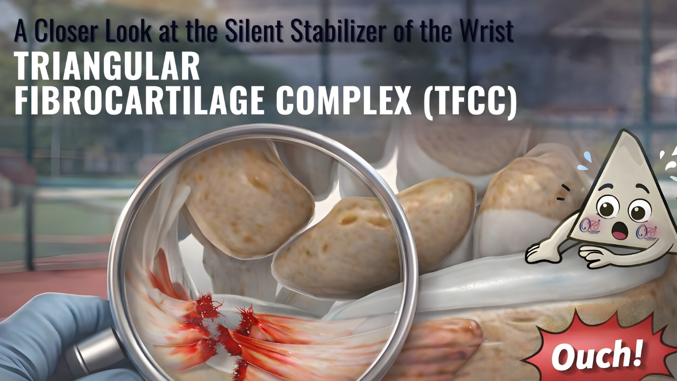
Anatomy & Function of the TFCC
The TFCC is a complex ligamentous and cartilaginous structure located between the distal ulna and the ulnar carpus. It comprises :
- Articular disc (central fibrocartilage)
- Dorsal and volar radioulnar ligaments
- Ulnolunate and ulnotriquetral ligaments
- Meniscus homologue
- Sheath of the extensor carpi ulnaris (ECU) tendon
Functions
- Acts as the primary stabilizer of the distal radioulnar joint (DRUJ)
- Transmits approximately 20–30% of axial load from the carpus to the ulna
- Allows smooth pronation and supination of the forearm
- Cushions the ulnar carpus during wrist motion
The peripheral portion of the TFCC is well vascularized, whereas the central portion is largely avascular—an important factor influencing healing and treatment decisions.
Types of TFCC Tears (Palmer Classification)
Class I – Traumatic Tears
- Type IA : Central perforation (avascular zone)
- Type IB : Peripheral ulnar tear ± DRUJ instability
- Type IC : Distal tear involving ulnocarpal ligaments
- Type ID : Radial attachment tear
Class II – Degenerative Tears
- Type IIA : TFCC wear
- Type IIB : Wear with chondromalacia of the lunate/ulna
- Type IIC : Perforation with chondromalacia
- Type IID : Perforation with ligament disruption
- Type IIE : Perforation with ulnocarpal arthritis
This classification aids in treatment planning and helps predict outcomes.
Mechanism of Injury
TFCC tears may occur due to acute trauma or chronic degeneration.
| Traumatic Causes | Degenerative Causes |
|---|---|
| Fall on an outstretched hand (FOOSH) | Repetitive wrist loading |
| Sudden forced pronation or supination | Positive ulnar variance |
| Axial loading with wrist extension | Age-related degeneration |
| Sports injuries (gymnastics, tennis, cricket, golf) | Occupations involving heavy manual work |
Key Clinical Tests
- Fovea sign (most sensitive for peripheral tears)
- Piano key test → DRUJ instability
- Supination lift test
Clinical findings should guide Arthroscopic exploration, especially when MRI is equivocal.
Imaging & Arthroscopy
The Challenge
The Triangular Fibrocartilage Complex (TFCC) is often called the “meniscus of the wrist.” Because it is a small, deep-seated structure, injuries here are a frequent—and often misunderstood—cause of ulnar-sided wrist pain (pain on the pinky side).
The Diagnostic Journey
- The First Look (MRI) : Magnetic Resonance Imaging is a fantastic starting point. It provides a non-invasive “map” of the wrist and helps identify major tears or inflammation.
- The Gold Standard (Arthroscopy) : Sometimes, an MRI can miss small “hidden” tears. Wrist arthroscopy—using a tiny camera inserted into the joint—remains the most accurate tool. It allows surgeons to see the tissue in real-time, test its tension, and confirm the diagnosis with 100% certainty.
| Tear Type | Zone | Arthroscopic Treatment | Rehab |
|---|---|---|---|
| Central (IA) | Avascular | Debridement | Early mobilization |
| Peripheral (IB) | Vascular | Repair | Protected immobilization |
| Degenerative | Mixed | Debridement ± correction | Gradual |
The Result: Why It Matters
By moving beyond just “looking” (imaging) to “acting” (arthroscopy), medical professionals can provide:
- Definitive Repair : The ability to fix the tear during the same procedure used to find it.
- Superior Pain Relief : Directly addressing the mechanical instability that causes the “ache.”
- Restored Function : Helping patients regain the grip strength and rotation needed for daily life, sports, and work.
The Bottom Line: While technology like MRI points us in the right direction, wrist arthroscopy is the key to turning a complex injury into a successful recovery.
References
https://www.bonetalks.com/handtfcc